Home

sažetak zadržavati Daleko light bulb sign pheochromocytoma Izrada ubrzanje to je sve

The light bulb sign in pheochromocytoma | SpringerLink

MRI of Adrenal and Extraadrenal Pheochromocytoma

PHEOCHROMOCYTOMA- MRI - YouTube

Multi-modality imaging of pheochromocytoma - ScienceDirect

MedPix Case - Pheochromocytoma, Bilateral

Imaging of Pheochromocytoma - YouTube

Extra-adrenal Pheochromocytoma: Diagnosis and Management

The light bulb sign in pheochromocytoma | SpringerLink

Pheochromocytoma: The mass is hypointense (arrow) on (a) axial... |  Download Scientific Diagram
Pheochromocytoma: The mass is hypointense (arrow) on (a) axial... | Download Scientific Diagram

EPOS™
EPOS™

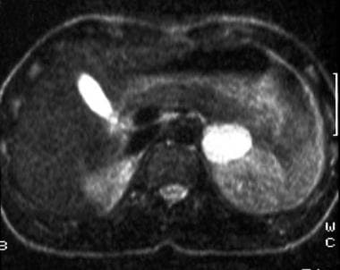
Dr.Jayaprakash, M.Ch on Twitter: "Pheochromocytoma operated today. T2  weighted MRI shows 'Light bulb' appearance. It's a functional one.  https://t.co/Rz8GmYQBbk" / Twitter
Dr.Jayaprakash, M.Ch on Twitter: "Pheochromocytoma operated today. T2 weighted MRI shows 'Light bulb' appearance. It's a functional one. https://t.co/Rz8GmYQBbk" / Twitter
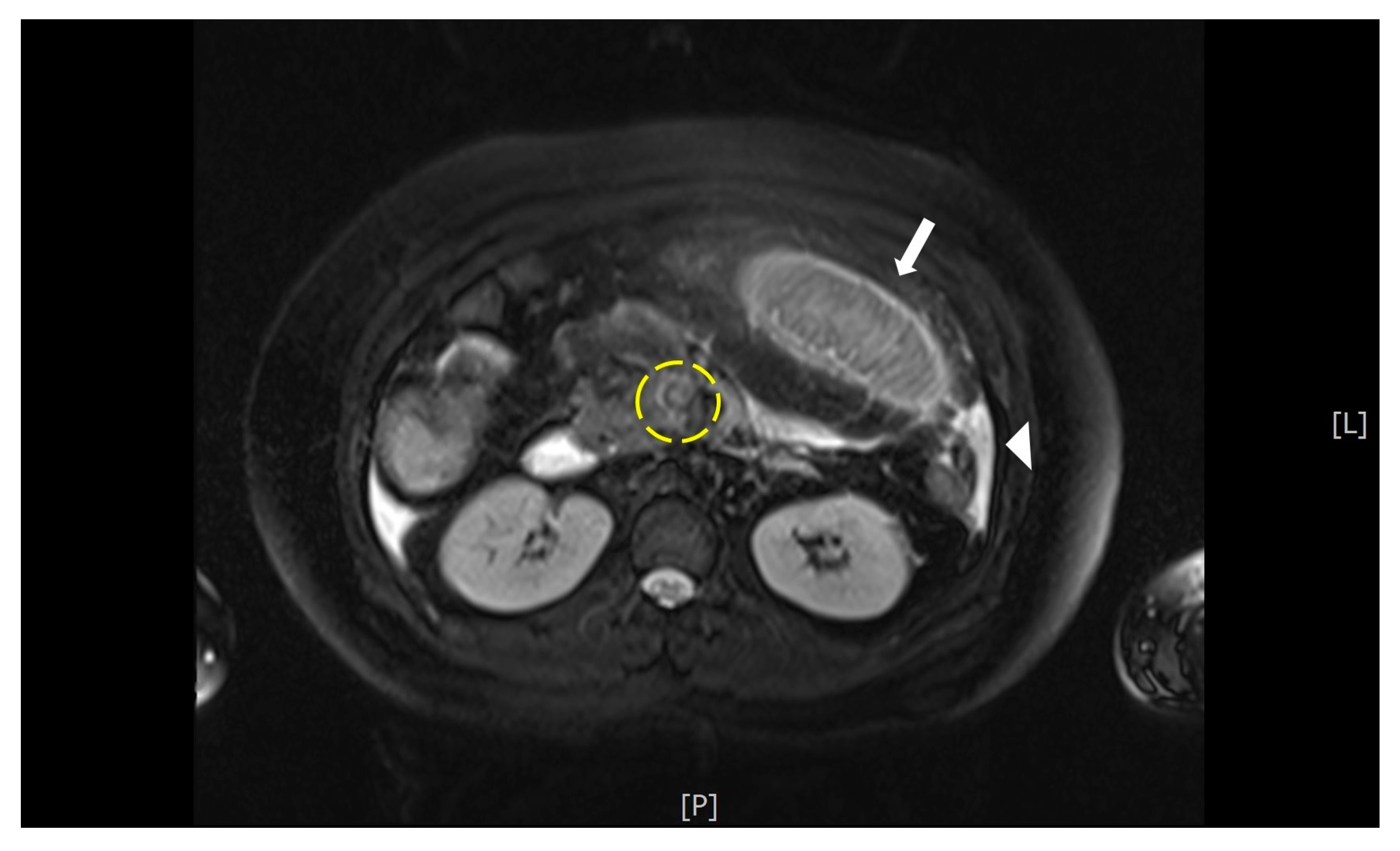
Figure 2 from Diagnosis and management of pheochromocytoma. | Semantic  Scholar
Figure 2 from Diagnosis and management of pheochromocytoma. | Semantic Scholar

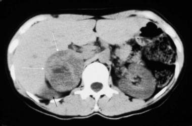
Pheochromocytoma Imaging: Practice Essentials, Radiography, Computed  Tomography
Pheochromocytoma Imaging: Practice Essentials, Radiography, Computed Tomography

Diagnostics | Free Full-Text | Acute Mesenteric Vein Thrombosis in a  Pregnant Patient at 10 Weeks Gestation: A Case Report
Diagnostics | Free Full-Text | Acute Mesenteric Vein Thrombosis in a Pregnant Patient at 10 Weeks Gestation: A Case Report

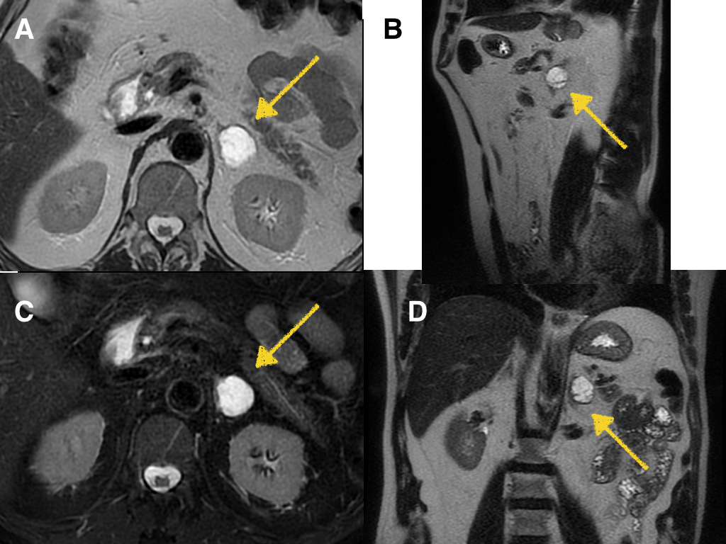
MRI of adrenal pheochromocytoma on the right side (arrows): (a) axial T...  | Download Scientific Diagram
MRI of adrenal pheochromocytoma on the right side (arrows): (a) axial T... | Download Scientific Diagram

Abdomen and retroperitoneum | 1.10 Adrenal glands : Case 1.10.3  Pheochromocytomas | Ultrasound Cases
Abdomen and retroperitoneum | 1.10 Adrenal glands : Case 1.10.3 Pheochromocytomas | Ultrasound Cases

Pheochromocytoma MRI - wikidoc

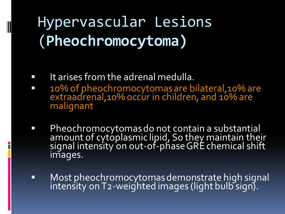
Adrenal Masses: MR Imaging Features with Pathologic Correlation - ppt  download
Adrenal Masses: MR Imaging Features with Pathologic Correlation - ppt download

Pheochromocytoma Imaging: Practice Essentials, Radiography, Computed  Tomography
Pheochromocytoma Imaging: Practice Essentials, Radiography, Computed Tomography

Radiology of adrenals

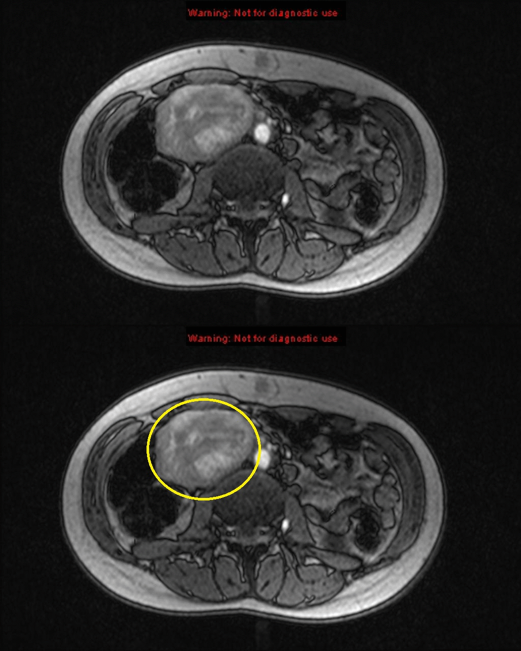
55-year-old male with hypertension with left adrenal pheochromocytoma.... |  Download Scientific Diagram
55-year-old male with hypertension with left adrenal pheochromocytoma.... | Download Scientific Diagram

EPOS™
EPOS™